TTE FOCUS Views Hack 3.0.0 + Redeem Codes
Developer: University Health Network
Category: Medical
Price: $4.99 (Download for free)
Version: 3.0.0
ID: org.ehealthinnovation.PIE.TTE-FOCUS
Screenshots


Description
In the practice of anesthesia, critical care and emergency medicine there is often a need for a quick, qualitative assessment of cardiac function. Focused Cardiac Ultrasound (FOCUS) is the process of carrying out this rapid qualitative assessment by practitioners in these fields. Training for carrying out a FOCUS assessment requires practitioners to understand the 3D structures of the heart that are seen in the 2D transthoracic echocardiography (TTE) image.
We have created this interactive application to assist with teaching and learning the assessment of normal and pathologic cardiac function with FOCUS. FOCUS assessments are based on a subset of 5 of the 20 standard TTE views:
Parasternal long axis
Parasternal short axis
Apical four chamber
Subcostal four chamber
Subcostal inferior vena cava
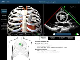
Users can view the TTE recordings for each of the 5 FOCUS views and see a corresponding 3D model of the probe, ultrasound plane, heart and rib cage for each view. Each FOCUS view can be selected from a menu at the left of the screen, or by using arrow buttons to go to the next or previous view in the list.
For each view, the 3D model of the probe, ultrasound plane, heart and rib cage can be rotated in the horizontal or vertical plane to view it from any angle. The rib cage can be removed, the part of the heart above the echo plane can be removed, and the heart model can be oriented so the structures correspond to the TTE image.
This module reviews the use of FOCUS to assess pericardial abnormalities of localised effusion and tamponade and their effect on cardiac function.
*NOTE: This app has not yet been updated to include the right ventricle, left ventricle and hypovolemic pathologies found on the website. http://pie.med.utoronto.ca/TTE/TTE_content/focus.html
Version history
3.0.0
2017-10-12
This app has been updated by Apple to display the Apple Watch app icon.
64bit compatible (compatible with iOS 11)
64bit compatible (compatible with iOS 11)
2.0
2014-01-15
**bug when updating to version 2.0. Please delete and then reinstall**
Layout has been updated.
Pericardial Pathology section added.
Layout has been updated.
Pericardial Pathology section added.
1.1
2013-10-09
Ways to hack TTE FOCUS Views
- Redeem codes (Get the Redeem codes)
Download hacked APK
Download TTE FOCUS Views MOD APK
Request a Hack
Ratings
3 out of 5
2 Ratings
